Moles

WHAT ARE  Moles? 

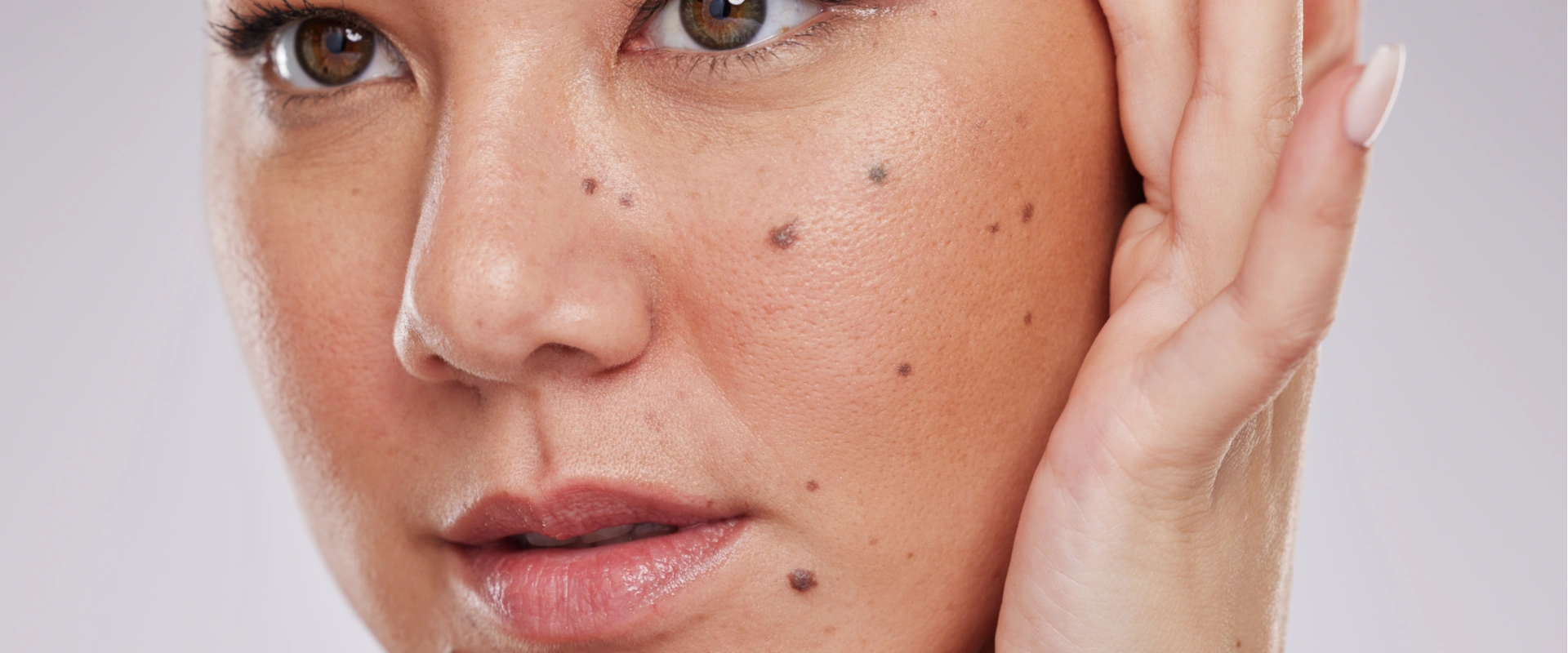
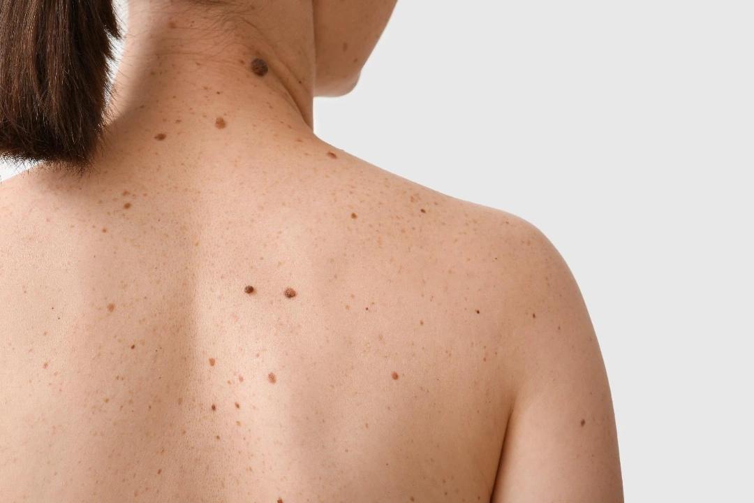
Moles, also known as nevi, are pigmented skin growths caused by clusters of melanocytes, which are pigment-producing cells. They may appear flat or raised and can vary in colour from light brown to nearly black. Moles are very common, and most adults have between 10 to 40 moles on their bodies.

Some moles are present from birth, while others appear gradually over time, often during childhood or adolescence. In most cases, moles are harmless and do not require treatment. However, changes in size, shape, or colour may indicate a risk of skin cancer and should be evaluated by a dermatologist.

moles skin cancer Singapore.
Moles are common pigmented spots on the skin, which are mostly benign and harmless. But roughly 25% of melanoma skin cancers originate from atypical moles.

What are the different types of  Moles? 

There are several different types of moles. Each with its distinct features, such as:
Type of MoleFeatures
Acquired Nevi
  • Develop over time, usually during childhood or adolescence
  • Most adults have 10 to 40 of these
Congenital Nevi
  • Present at birth
  • Larger congenital moles carry a higher risk of developing into melanoma later in life
Compound Nevi
  • Raised and often darker
  • Melanocytes are found in both the dermis and epidermis
Intradermal Nevi
  • Typically skin-coloured and raised
  • Located deeper in the skin
Junctional Nevi
  • Flat, brown moles located at the junction between the top and middle layers of the skin
Dysplastic (Atypical) Nevi
  • Irregular in shape or colour and may resemble melanoma
  • These moles carry a higher risk of becoming cancerous and require closer monitoring

What causes Moles?

The exact cause of mole formation is not fully understood. But genetics and environmental factors, particularly sun exposure, play a role. Some of the key contributing factors include:

  • Genetic predisposition – if your parents or siblings have numerous or atypical moles, you are more likely to develop them too.
  • Hormonal changes – moles may darken or appear during puberty, pregnancy, or hormonal treatments due to increased melanin activity.
  • Immune and cellular factors – some immune conditions and skin disorders may influence how melanocytes behave.
  • Skin type – individuals with fair skin, light hair, and light-coloured eyes are more prone to mole formation.
  • Sun exposure – ultraviolet (UV) rays from the sun or tanning beds stimulate melanocyte activity, leading to the development of new moles and changes in existing ones.

What are the characteristics of Moles?

Benign moles typically have a consistent and predictable appearance. As such, knowing what normal moles look like helps you detect any changes that may warrant medical attention.

These characteristics include, but are not limited to:

  • Size – most harmless moles are smaller than 6 mm in diameter, about the size of a pencil eraser. Larger moles, especially if growing, should be examined.
  • Symmetry – a healthy mole is usually round or oval with both halves looking similar. Asymmetry, where one side does not mirror the other, may be a warning sign.
  • Stable appearance – benign moles remain consistent in shape, colour, and size over time. Any noticeable evolution could indicate an abnormality.
  • Uniform colour – normal moles are typically a single shade of brown, tan, or black. Moles that contain multiple colours, including red, white, blue, or grey, require closer inspection.
  • Well-defined borders – benign moles tend to have smooth, clearly defined edges. In contrast, moles with blurry, jagged, or notched borders may indicate atypia.

Who is at risk of developing Moles?

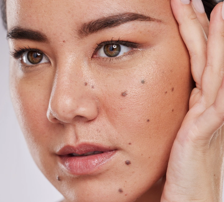
Although moles can form in anyone, there are certain individuals who are more predisposed due to several factors. The groups that are at higher risk are:

  • Family history of moles or melanoma – genetics play a strong role. If close relatives have numerous or atypical moles, your risk is higher.
  • Hormonal changes – puberty, pregnancy, and hormonal therapy can increase melanin activity, causing new moles to appear or existing ones to darken.
  • Weakened immune system – those with autoimmune conditions, organ transplants, or certain medications may have altered skin cell regulation, increasing mole activity.
  • Fair-skinned individuals – those with lighter skin tones, blue or green eyes, and blonde or red hair produce less melanin and are more vulnerable to UV-related skin damage.
  • Frequent sun exposure – living in sunny climates like Singapore or spending lots of time outdoors without sun protection can stimulate melanocyte activity, leading to more mole development.
  • Excessive use of tanning beds – artificial UV radiation is just as damaging as natural sunlight and significantly raises the risk of both mole formation and skin cancer.
what increases mole development.
Prolonged sun exposure stimulates melanin activity, increasing the likelihood of new mole formation, especially in fair skin types.

How does a Mole become cancerous?

Moles consist of melanocytes, the same cells involved in melanoma. Although most moles remain stable, a small number may undergo changes that lead to uncontrolled cell growth.

Melanoma arises when melanocytes become cancerous and begin to multiply abnormally. This can occur within an existing mole or develop as a new lesion.

Some of the triggers for mole-to-melanoma transformation may include:

  • Chronic irritation or trauma – repeated friction, rubbing, or injury to a mole may stimulate abnormal changes, although this is less common.
  • Genetic mutations – certain inherited or acquired mutations may disrupt normal cell growth controls.
  • UV damage – excessive ultraviolet exposure from the sun or tanning beds damages DNA and increases the risk of abnormal melanocyte behaviour.
  • Weakened immune defences – a suppressed immune system may fail to detect and remove abnormal cells early on.


About 25% to 30% of melanomas arise from pre-existing moles, with the rest appearing as new lesions. This is why routine monitoring and early intervention are critical.

What signs indicate that a Mole is skin cancer?

Most moles are completely harmless and do not change throughout your lifetime. However, certain signs may suggest that a mole is becoming atypical or possibly cancerous.

You should consult a dermatologist if a mole:

  • Becomes itchy, painful, or starts to bleed
  • Develops a crusty, scaly, or rough texture
  • Has a blurry, irregular, or poorly defined border
  • It is larger than 6 mm (about the width of a pencil eraser)
  • It starts to change rapidly in size, shape, or colour without a clear cause
  • Looks different from your other moles, known as the “ugly duckling” sign

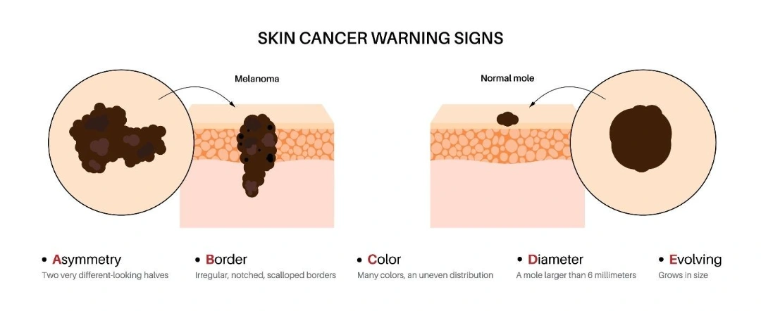
The ABCDE rule is a simple but effective tool for identifying potentially suspicious moles:

  • A: Asymmetry – one half of the mole does not match the other
  • B: Border – the edges are irregular, jagged, or poorly defined
  • C: Colour – uneven tones or multiple colours present
  • D: Diameter – larger than 5 to 6 mm
  • E: Evolution – changes in any aspect, such as size, colour, height, sensation
signs of cancerous moles.
Look out for changes in asymmetry, border, colour, diameter, and evolution in your moles, and early detection can save lives.

How are Moles diagnosed in Singapore?

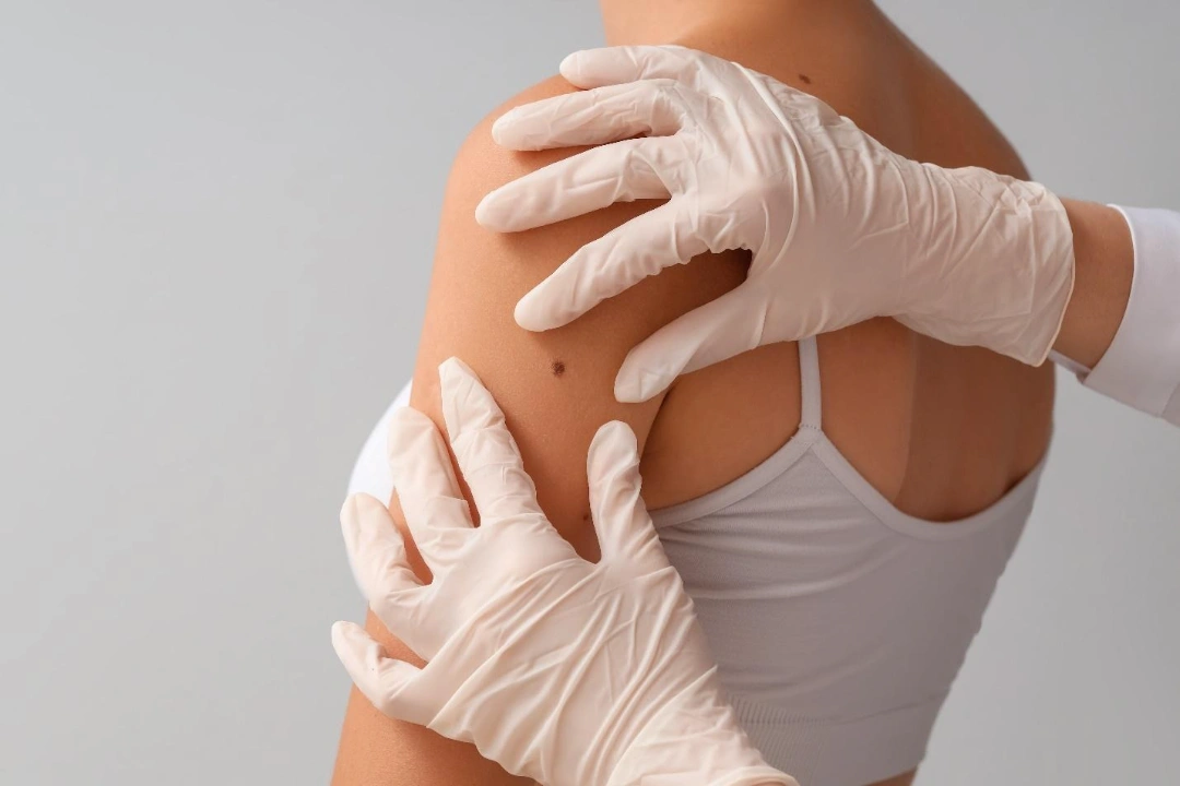
When it comes to diagnosing moles, we use a structured and meticulous approach to accurately assess and diagnose moles. Our goal is to differentiate between harmless and suspicious lesions to ensure safe and timely treatment.

Our diagnostic process includes:

  • Comprehensive skin examination our dermatologist visually assesses the mole’s size, colour, texture, and shape. This includes checking for asymmetry, border irregularity, and other warning signs. A full-body mole check may be recommended for patients with numerous moles.
  • Medical history and symptom review you will be asked about any recent changes, family history of skin cancer, sun exposure habits, or personal risk factors.
  • Dermoscopy a non-invasive tool that provides a magnified view of the mole’s internal structure and pigmentation patterns. This helps distinguish benign moles from atypical or malignant ones.
  • Skin biopsy if the mole appears suspicious, a biopsy may be performed to confirm the diagnosis. This involves removing a small tissue sample and sending it to a laboratory for histological examination. Some of the potential methods include:
    • Excision biopsy the entire mole is removed with a scalpel
    • Punch biopsy a circular blade removes a deeper skin core
    • Shave biopsy surface layers of the mole are shaved off

When is Mole removal recommended?

Mole removal may be suggested for medical, aesthetic, or practical reasons. Even benign moles can sometimes cause discomfort or interfere with daily life.

Some of the common reasons for mole removal include:

  • Aesthetic concerns – many patients choose to remove facial or visible moles for cosmetic reasons
  • Irritation and friction – moles that catch on clothing, jewellery, or shaving razors can become inflamed or painful
  • Personal preference – some individuals opt for removal to eliminate worry, even if the mole is harmless
  • Suspicion of malignancy – moles that are evolving or show signs of atypia should be removed and examined under a microscope

Before any removal, our dermatologist will perform a thorough assessment to ensure the right technique is chosen based on the mole's type, depth, and location.

why consider mole removal.
Mole removal can be done for both medical and cosmetic reasons.

How are Moles removed?

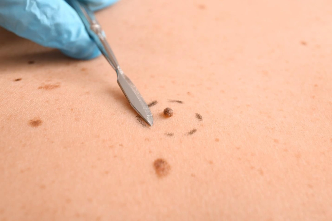
At Cove Aesthetic Clinic, mole removal is a safe and straightforward outpatient procedure performed by trained medical professionals. The appropriate method depends on the mole’s type, size, depth, location, and whether it shows any signs of potential malignancy.

Each removal technique is selected carefully to balance clinical effectiveness, aesthetic outcomes, and, when needed, histological assessment.

This is the most comprehensive method and is often used for moles that are deep, large, or clinically suspicious.

  • The entire mole, along with a small margin of surrounding skin, is removed using a scalpel.
  • The area is then closed with stitches to aid healing and reduce scarring.
  • The excised tissue is typically sent to a laboratory for histopathological analysis.
  • The treatment is suitable for suspicious, deep, or larger moles where malignancy needs to be ruled out.

A common method for removing raised or protruding moles that are benign in appearance.

  • A surgical blade is used to shave off the mole flush with the skin’s surface.
  • Stitches are generally not required.
  • Local anaesthesia is applied to ensure the procedure is comfortable.
  • Healing typically results in a flat, skin-coloured mark or slight pigment difference.

This involves the use of a small, circular tool to remove both the mole and a column of surrounding tissue.

  • Typically used for smaller, deeper moles.
  • Local anaesthetic is administered before the procedure.
  • A few stitches may be required to close the site.
  • Tissue is sent for lab analysis if needed.

Laser mole removal is a non-invasive option for moles that are flat, superficial, and benign.

  • A targeted laser, such as a CO2 or Q-switched laser, breaks down the mole’s pigment, causing it to fade over time.
  • No cutting or stitching is required.
  • Typically done over one or more sessions, depending on the mole’s response.
  • Not suitable for moles suspected to be cancerous, as no tissue is collected for biopsy.

This method involves freezing the mole using liquid nitrogen, which destroys the pigmented cells.

  • The treated tissue will scab and fall off naturally within a few days.
  • Best suited for small, benign, and superficial moles.
  • Not commonly used for deeper or suspicious lesions.

Electrocautery uses heat generated by an electric current to burn and destroy mole tissue.

  • Often used for small or superficial benign moles.
  • Provides excellent control over bleeding during removal.
  • May be combined with curettage (scraping) for more thorough results.
benefits of mole removal.
There are several safe and effective ways to remove moles, with each method selected based on the mole’s type, size, and location to ensure optimal medical and cosmetic outcomes.

Summary

Whether you are concerned about a changing mole or simply wish to remove one for cosmetic reasons, expert evaluation is the first step. At Cove Aesthetic Clinic, our experienced team offers safe, effective mole assessment and removal tailored to your needs, with a focus on both medical accuracy and aesthetic outcomes.

Book a consultation with us today to have your moles assessed and explore the most suitable treatment options for your skin.

Frequently Asked Questions

No. Most moles are benign and do not pose a health risk. However, certain types, such as dysplastic or atypical moles, have a higher chance of becoming cancerous and should be monitored regularly.

Only a medical professional can confirm whether a mole is cancerous, usually through a biopsy. However, signs such as changes in size, shape, colour, bleeding, or itching may warrant immediate medical attention. The ABCDE rule is a helpful guide.

New moles can form due to hormonal changes, sun exposure, or genetics. If a new mole appears and looks unusual or changes rapidly, it is best to have it evaluated.

Yes, hormonal changes during pregnancy can cause existing moles to darken or new ones to form. These changes are usually benign, but it is best to have any rapidly changing moles checked by a dermatologist.

You may not be able to prevent all moles, especially if you are genetically predisposed. However, limiting UV exposure, wearing sunscreen daily, and avoiding tanning beds can help reduce the development of new moles.

No, mole removal should never be attempted at home. DIY methods can lead to incomplete removal, infections, scarring, or worse—missed diagnoses of skin cancer. Always seek professional evaluation and removal from a trained medical provider.

Mole removal procedures are generally not painful. Local anaesthesia is applied to numb the area before removal. You may feel mild pressure during the procedure, and some tenderness or redness afterwards, which typically resolves within a few days.

Some scarring is possible, depending on the mole’s size, depth, and the removal method used. However, our dermatologists take great care to minimise scarring, especially for facial or visible areas. Over time, most scars fade significantly.

Laser removal is safe and effective for treating small, flat, non-cancerous moles. However, it is not recommended for suspicious moles, as no tissue is left behind for biopsy. Our dermatologist will determine if it is appropriate for your case.

In some cases, yes. If a mole is not completely removed, especially with superficial methods like shaving, it may regrow. Complete surgical excision reduces the likelihood of recurrence.

Yes. Many patients choose to remove moles that are visible, raised, or bothersome. Cosmetic mole removal is safe when performed by a qualified medical professional and typically leaves minimal scarring.

Yes. Mole removal is safe for children and teens when medically indicated or requested for cosmetic reasons. Evaluation by a dermatologist is essential before proceeding with removal in younger patients.

Healing times vary depending on the technique used and the individual’s skin type. In most cases, surface healing takes 7 to 10 days. Complete healing and scar fading can take several weeks to a few months.

NO TWO FACES ARE THE SAME, AND NEITHER ARE our treatments.

Customised treatment plans always work best. We tailor each treatment plan to your specific needs, ensuring you get the results you desire.  Our patient’s journey starts before their booking – and continues after their COVE experience.
Book an appointment
STEP ONE

Book a Consultation

We require new patients to attend a consultation with our doctors so we can better understand your aesthetic concerns. At Cove, we genuinely care for our patients. Consultations are in-depth, never rushed, and we will never recommend treatments that you do not need. Simply drop us a WhatsApp or a phone call to book an appointment!
STEP TWO

Come On Over

It’s time to come into COVE and meet us for your consultation! During this phase, we encourage you to maintain an open mind. While you may have a vision in mind, our doctors may offer different advice based on their assessment of your condition and desired treatment outcomes.
STEP THREE

Begin Your Transformation

You’ve consulted our Cove Experts, and the treatment plan has been discussed with you. We’re now ready to make your transformation happen! To ensure no time is wasted, we recommend starting your treatment in the same session as your consultation. You’ll love how each visit to COVE is a boost to your self-confidence! 

View More Concerns

Wheelock Place | 501 Orchard Rd, #04-07, Singapore 238880
Guoco Tower  | 7 Wallich Street, #B1-14, Singapore 078884

WHEELOCK PLACE (ORCHARD)

501 Orchard Rd, #04-07, Singapore 238880
hello@coveaesthetics.com.sg
(65) 6734 3977
(65) 8082 0998
Mon-Fri: 10:00am to 8:00pm

Sat: 9:30am to 4:00pm 

Closed on Sundays & PH

GUOCO TOWER (TANJONG PAGAR)

7 Wallich Street, #B1-14, Singapore 078884
hello@coveaesthetics.com.sg
(65) 8339 3518
(65) 8339 3518
Mon-Fri: 10:00am to 8:00pm

Sat: 9:30am to 4:00pm 

Closed on Sundays & PH
Copyright © 2026 Cove Aesthetic Clinic. All Rights Reserved.
Copyright © 2026 Cove Aesthetic Clinic.
All Rights Reserved.
chevron-down